PRK Surgery: Why and When is it Indicated?
PRK (Photorefractive Keratectomy) is one of the foundational refractive procedures used to correct myopia (nearsightedness), hyperopia (farsightedness), and astigmatism. Although the visual recovery period is longer than that of LASIK, PRK is widely performed and offers distinct advantages in specific clinical profiles. The underlying optical mechanism is identical to LASIK: an excimer laser reshapes the anterior corneal surface so incoming light focuses precisely on the retina, restoring clear uncorrected vision.
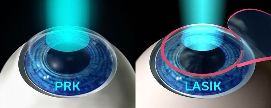
The fundamental difference between PRK and LASIK lies in how the treatment zone is accessed. In LASIK, a thin surface flap is created and folded back to expose the corneal stroma. In PRK, the superficial corneal epithelium is completely removed rather than folded. In both techniques, an excimer laser ablates the stromal bed to correct refractive errors.
Another variation of surface ablation is LASEK (Laser-Assisted Sub-Epithelial Keratectomy). In LASEK, the epithelium is loosened (typically using a diluted alcohol solution within a trephine), the stromal bed is sculpted with the excimer laser as in PRK, and the epithelial sheet is repositioned over the treatment zone. Compared to standard PRK, LASEK can exhibit a prolonged visual recovery because biological reattachment of the manipulated epithelium often takes longer than fresh epithelial regrowth.
PRK vs. LASIK
Long-term visual outcomes of PRK and LASIK are comparable. However, early recovery after PRK requires more time due to the cellular regeneration of the epithelium. Additionally, temporary risks of post-operative haze and ocular infection are slightly higher during initial epithelial closure.
Despite these factors, PRK eliminates flap-related complications and preserves a greater stromal bed. Because no stromal tissue is consumed by a flap, the entire corneal thickness remains available for treatment. Consequently, PRK is the preferred option for patients with thin corneas, anterior basement membrane disorders, high-impact athletic lifestyles, or those undergoing enhancement after previous LASIK. Furthermore, the risk of corneal ectasia is minimized.
How is PRK Performed?
In PRK, the central corneal epithelium is gently removed using a diluted alcohol solution, mechanical debridement, or an automated brush. The excimer laser then reshapes the stromal curvature with sub-micron precision. Finally, a therapeutic soft bandage contact lens is placed on the eye. New epithelial cells regenerate completely beneath the lens within 3 to 5 days, after which the surgeon removes the bandage lens.
Perioperative Instructions
Pre-Operative:
Consult a subspecialist for a comprehensive diagnostic evaluation, including:
Pupil size measurement (pupillometry)
Tear film analysis and dry eye assessment
Corneal topography and tomography (curvature evaluation)
Corneal pachymetry (thickness measurement)
Your surgeon will also review your systemic medical history and medications. Contact lens wear must be discontinued prior to evaluation, as lenses alter baseline corneal geometry.
Intraoperative:
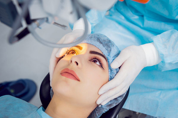
PRK is an outpatient procedure lasting approximately 15 minutes. A mild oral sedative may be administered for relaxation alongside topical anesthetic eye drops. An eyelid speculum prevents blinking. The patient fixates steadily on a target light while the surgeon delivers computer-guided laser pulses under surgical biomicroscopy. The laser sculpts corneal tissue at the microscopic level without sharp pain (though mild intraocular pressure sensations may occur). Both eyes are typically treated consecutively on the same day.
Immediate Post-Operative Care:
Following a brief rest period, the patient is discharged (an adult escort is required). Prescribed antibiotic, anti-inflammatory, and analgesic eye drops are initiated to manage post-procedural discomfort and support healing. Adherence to postoperative visits over the subsequent weeks is essential.
Given the extended healing curve of surface ablation, functional vision for driving is generally achieved within 1 to 3 weeks, with complete visual crispness and stability settling within 3 to 6 months