
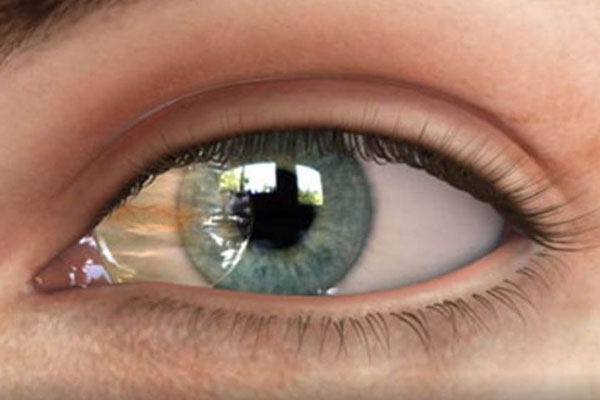
Pterygium Disease and Its Surgery
Pterygium and pinguecula are commonly seen in individuals who spend long hours outdoors without sunglasses. Even children are not exempt from this condition.
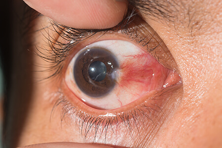
In affected individuals, a fleshy, pinkish growth appears on the white sclera/conjunctiva. This growth most frequently originates on the side closest to the nose and extends toward the center of the eye. Pterygium is a benign (non-cancerous) growth that typically develops slowly, and its progression may even halt temporarily. In advanced cases, it can enlarge sufficiently to cover the pupil, leading to visual impairment.
Pterygium can affect both eyes. While generally not dangerous, it often causes a persistent foreign body sensation. When inflamed, vascularized, and red, medical intervention becomes necessary.
Symptoms
Pterygium often produces no symptoms other than its visible appearance. However, marked growth can trigger redness and inflammation. As it extends onto the cornea, it distorts its curvature—inducing astigmatism and blurred vision.
Common symptoms include:
Burning sensation
Gritty sensation (feeling of sand in the eye)
Itching
Foreign body sensation
Blurred vision
Causes of Pterygium
Leading ophthalmic consensus identifies key causative factors as:
Prolonged exposure to ultraviolet (UV) sunlight
Dry eye syndrome
Chronic ocular irritation from wind and dust
Prevalence is exceptionally high in equatorial and sunny climates. It predominantly presents in adults aged 20 to 40, with a higher incidence among men.
Pterygium is closely related to pinguecula—a benign, yellowish conjunctival nodule adjacent to the cornea sharing identical etiologies, especially UV exposure. Because pinguecula disrupts the uniform spread of the tear film, it frequently induces ocular discomfort, dryness, and occasional redness.
Treatment Methods
If you suspect you have pterygium, consult an ophthalmologist for a comprehensive slit-lamp biomicroscopy examination. Mild, asymptomatic cases do not require intervention. Medical therapy is indicated when ocular surface inflammation and hyperemia develop:
Lubricating eye drops and ointments (such as Blink)
Vasoconstrictor eye drops (such as Naphcon-A)
Short-term topical corticosteroids (such as FML or Lotemax)
When pterygium causes persistent irritation, astigmatism, or visual obstruction, surgical excision is performed as an outpatient procedure. Indications for surgery include:
Failure of conservative medical therapy
Threat of visual axis involvement or vision loss
Persistent cosmetic distress and chronic redness
Surgical removal combined with conjunctival autografting (using the patient’s own conjunctiva) or amniotic membrane transplantation (AMT) is used to reconstruct the bare scleral bed. The procedure takes approximately 30 to 45 minutes. An eye patch is worn for 1 to 2 days postoperatively. Patients can resume routine daily activities within a few days (avoiding swimming and eye rubbing). Topical corticosteroid drops are prescribed for several weeks or months to suppress inflammation and minimize recurrence risk.
Recurrence remains the primary surgical complication. Conjunctival autografts and amniotic membrane grafts significantly reduce recurrence rates from up to 50% to roughly 5–10%. Other potential surgical risks include induced astigmatism, corneal scarring, or scleral thinning. Because over 97% of recurrences occur within the first 12 months, post-operative clinical follow-up is recommended for one full year.
Prevention of Pterygium
Daily use of sunglasses blocking 99% to 100% of UVA and UVB radiation is the most effective preventive measure, even on overcast days. Wraparound frames offer optimal protection against UV rays, wind, and airborne debris. Sunglasses are also strongly advised during driving, and lubricating artificial tears help maintain ocular surface hydration in dry environments.